
ACCESS:
Please contact the CMIF staff before bringing samples to the lab. Before beginning your TEM project, please email the CMIF staff with a detailed description of your experiment. We will be able to provide you with advice on how to prepare your samples as well as provide the appropriate fixative in a timely manner. If you are not familiar with the complexities of a TEM project, we strongly suggest that you meet directly with a CMIF staff member to discuss your project in detail. Sample processing for TEM is by appointment only and we require 2-3 weeks notice prior to processing samples. Please consider this when planning your experiments since processing delays will result in degradation of the sample and less than optimal images.
RATES:
Please refer to our rates page.
INFORMATION:
This facility is proficient in the examination of biological specimens. For imaging and characterization of materials (i.e. polymers, nanomaterials), we refer you to the OSU Center for Electron Microscopy and AnalysiS.
Provided sufficient lead time, services provided by the CMIF staff can be tailored to the needs of the investigator. If the project warrants extensive use of the TEM, we provide individual training for independent microscope use. However, if the project requires limited TEM use, it will be more cost effective to have the CMIF Electron Microscopist obtain images for you. In addition to instrument instruction, the CMIF provides the following services: sample fixation, embedding, negative staining and the production of semi- or ultra-thin sections, and cryo-sectioning.
The FEI Tecnai G2 Spirit TEM is ideal for imaging all biological specimens, including ultra-thin sections and negatively stained samples. This instrument has a 20-120 KV range and is capable of producing high resolution, digital images. Read more about the capabilities of the Tecnai G2 Spirit here.
Auxiliary equipment for specimen preparation includes a Pelco Biowave microwave tissue processing system.
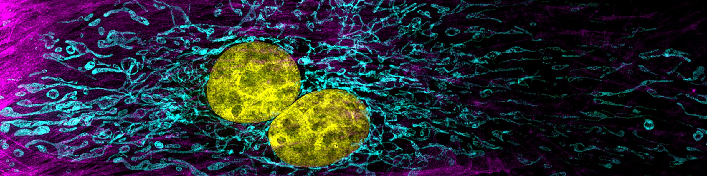
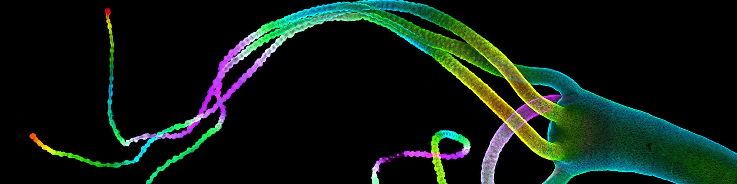
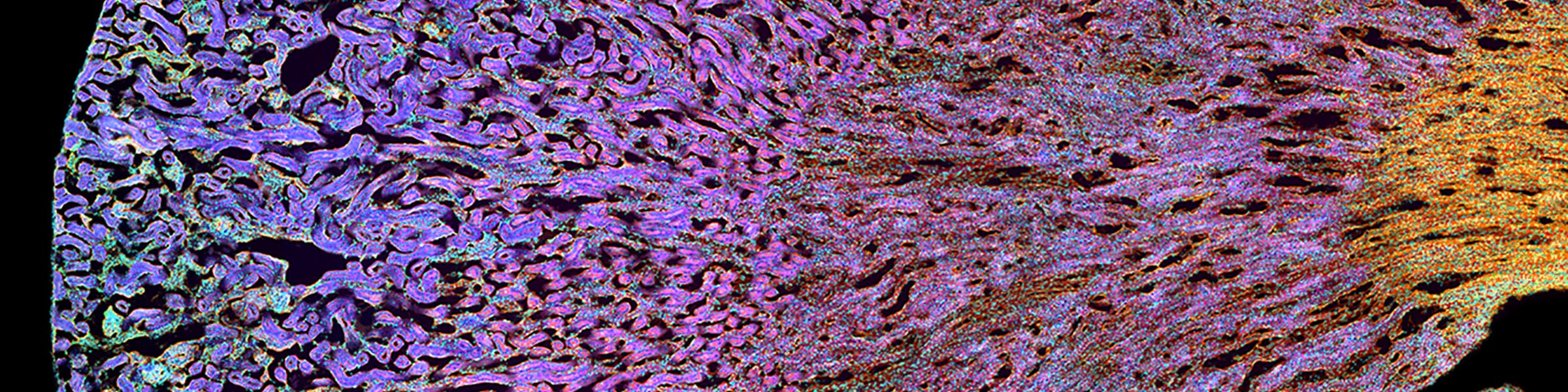
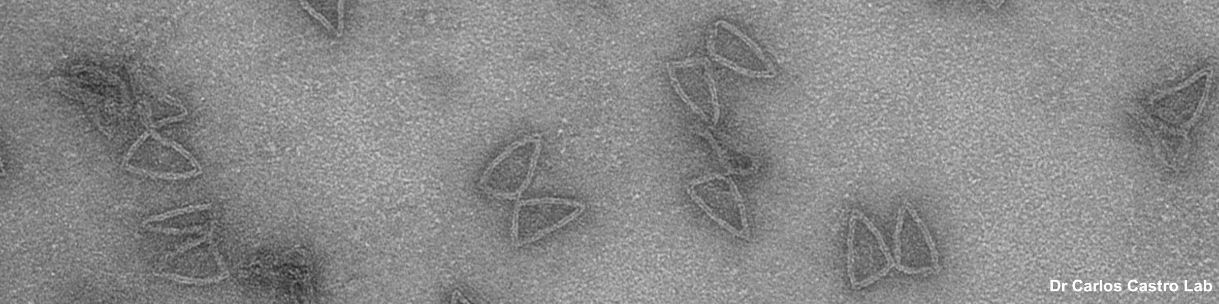
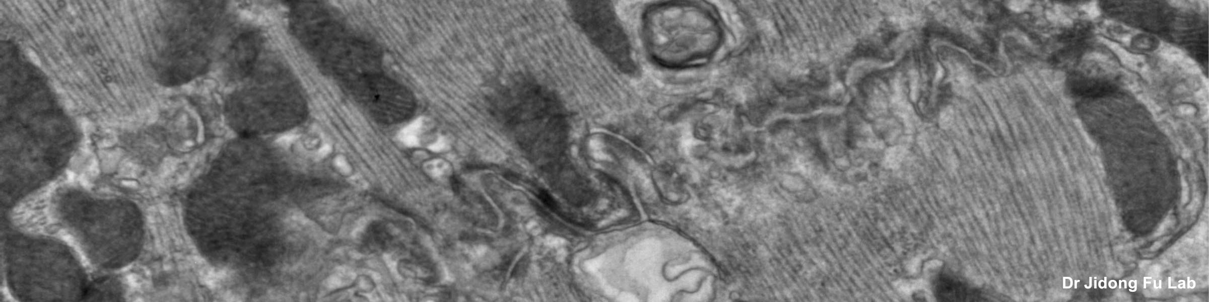
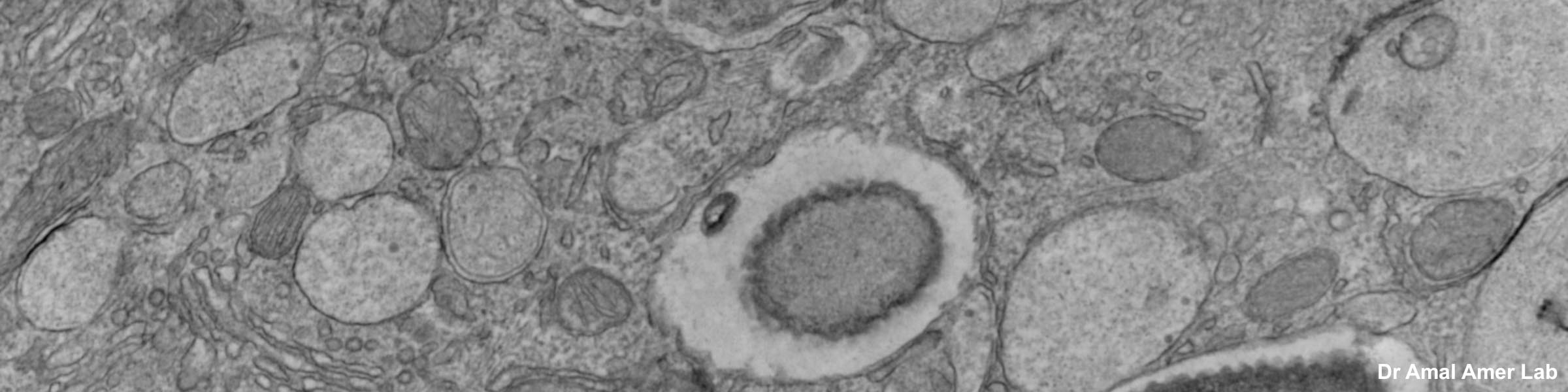
SAMPLE IMAGES:
Below are some images representing the services we provide.
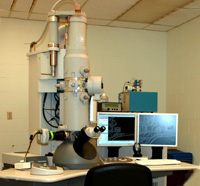 FEI Tecnai G2 Spirit TEM
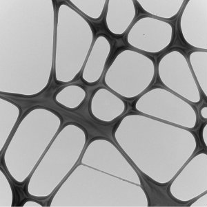 Brightfield Image of substrate
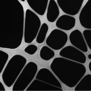 Darkfield Image of substrate
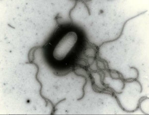 NEGATIVE STAINING Salmonella
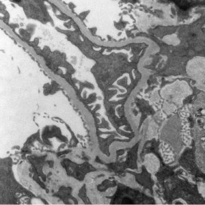 TISSUES Kidney Glomerulus
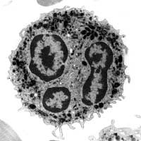 CULTURED CELLS neutrophil