ACCESS:
The Olympus FV1000 Multiphoton is available for independent use. Complete confocal microscope (single photon) training is required with NO exception. Training is performed on an individual basis using the researchers samples and instrument rates and technical assistance fees apply during individual instruction.
BSL-2 training from OSU EHS is required for all researchers wishing to perform live cell imaging at the CMIF. A signed form must be submitted to the facility before the start of any live cell imaging project.
RATES:
Please refer to our rates page.
INFORMATION:
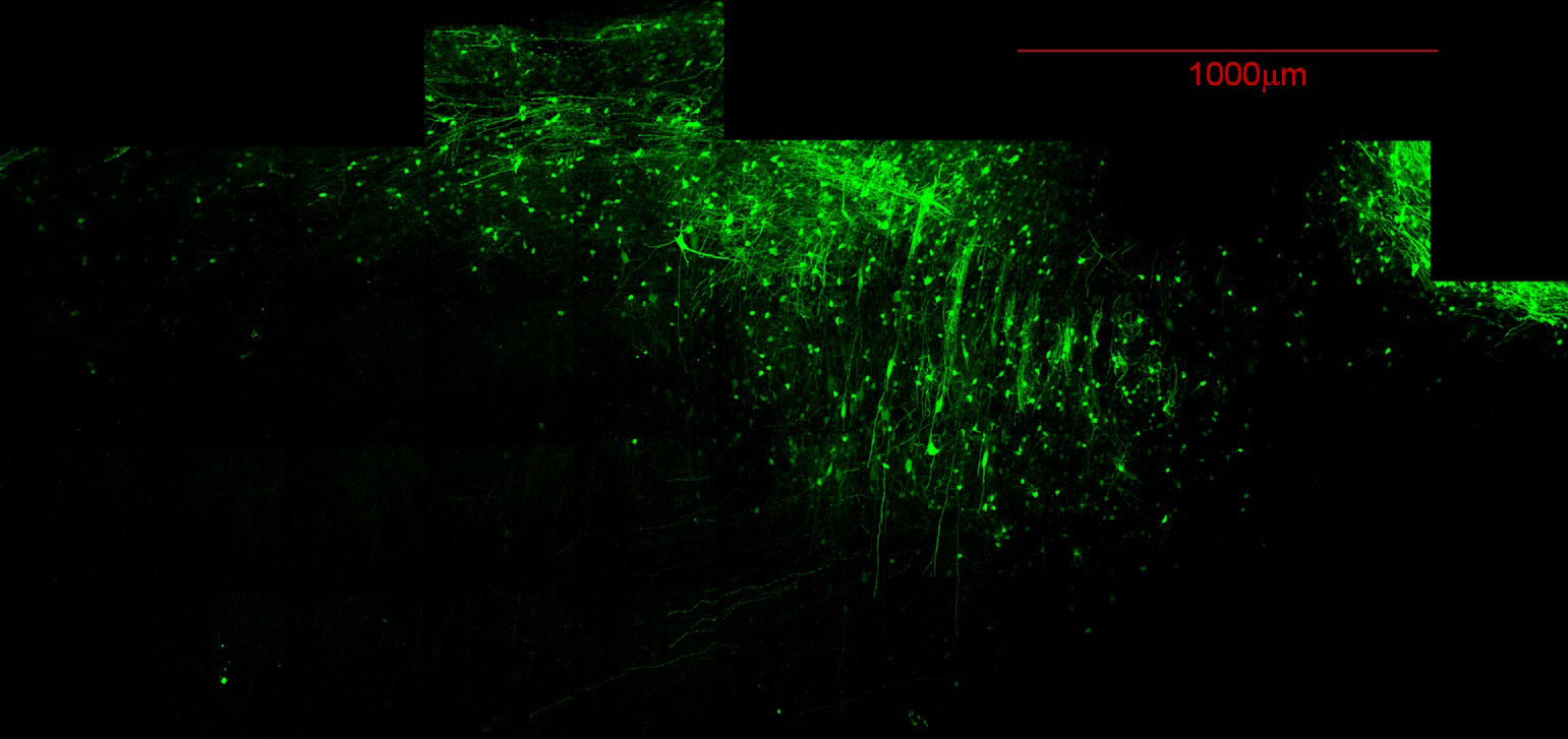
The Olympus FV1000 MPE Multiphoton Laser Scanning Confocal is equipped with a DeepSee MaiTai titanium -sapphire laser, a 25x1.05 N.A. 2mm working distance water immersion objective lens, four non-descanned detectors and a forward, second harmonic generation (SHG) detector. SHG can image collagen, myosin, and some polysaccharides such as cellulose and starch without staining. This microscope enables researchers to image deep into fixed or living tissues since the infrared light can penetrate up to 1 millimeter into the sample. Several support instruments are available for intravital, live cell or live tissue imaging including a temperature controlled imaging chamber and a rodent oximeter. A vibratome is available for thick sectioning of fixed tissues.
This system also functions as a standard confocal microscope system with 405,488,594,633 nm excitation lasers, 3 fluorescence detectors and a transmitted detector for DIC imaging. A motorized stage is available for capturing tiled images or tiled image stacks. Additional water immersion objective lenses are available for use with the Multiphoton including a 40X lens and a 100X lens. Dry and oil objective lenses can also be used on this system.
SAMPLE IMAGES:


