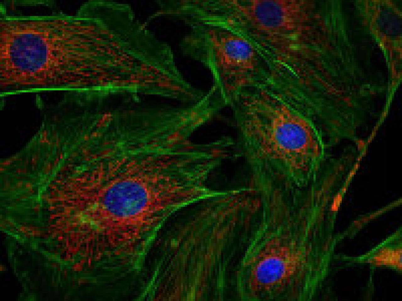
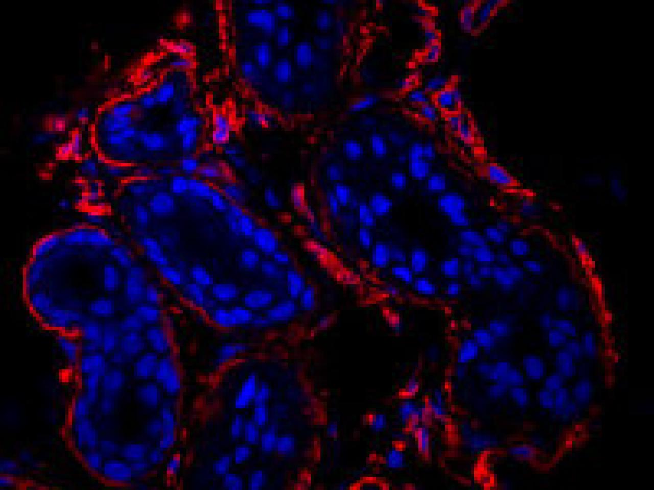
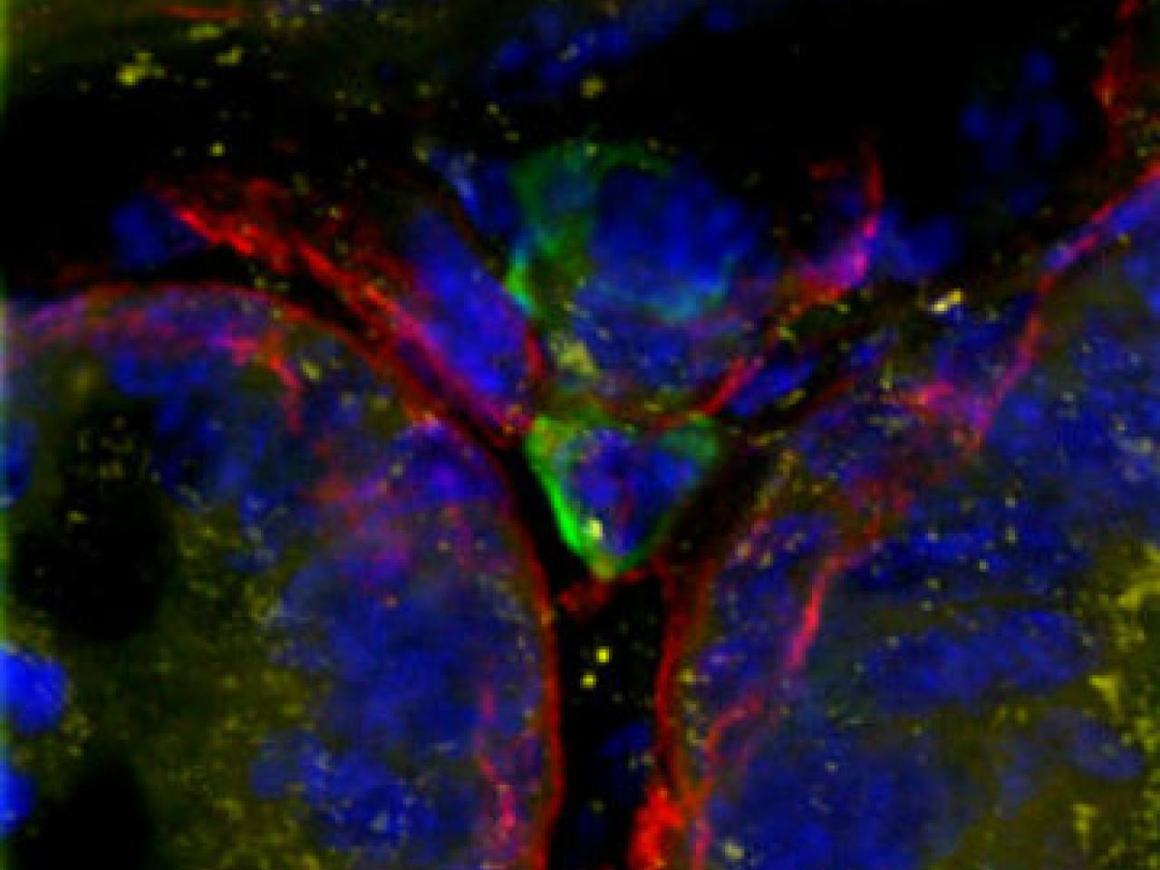
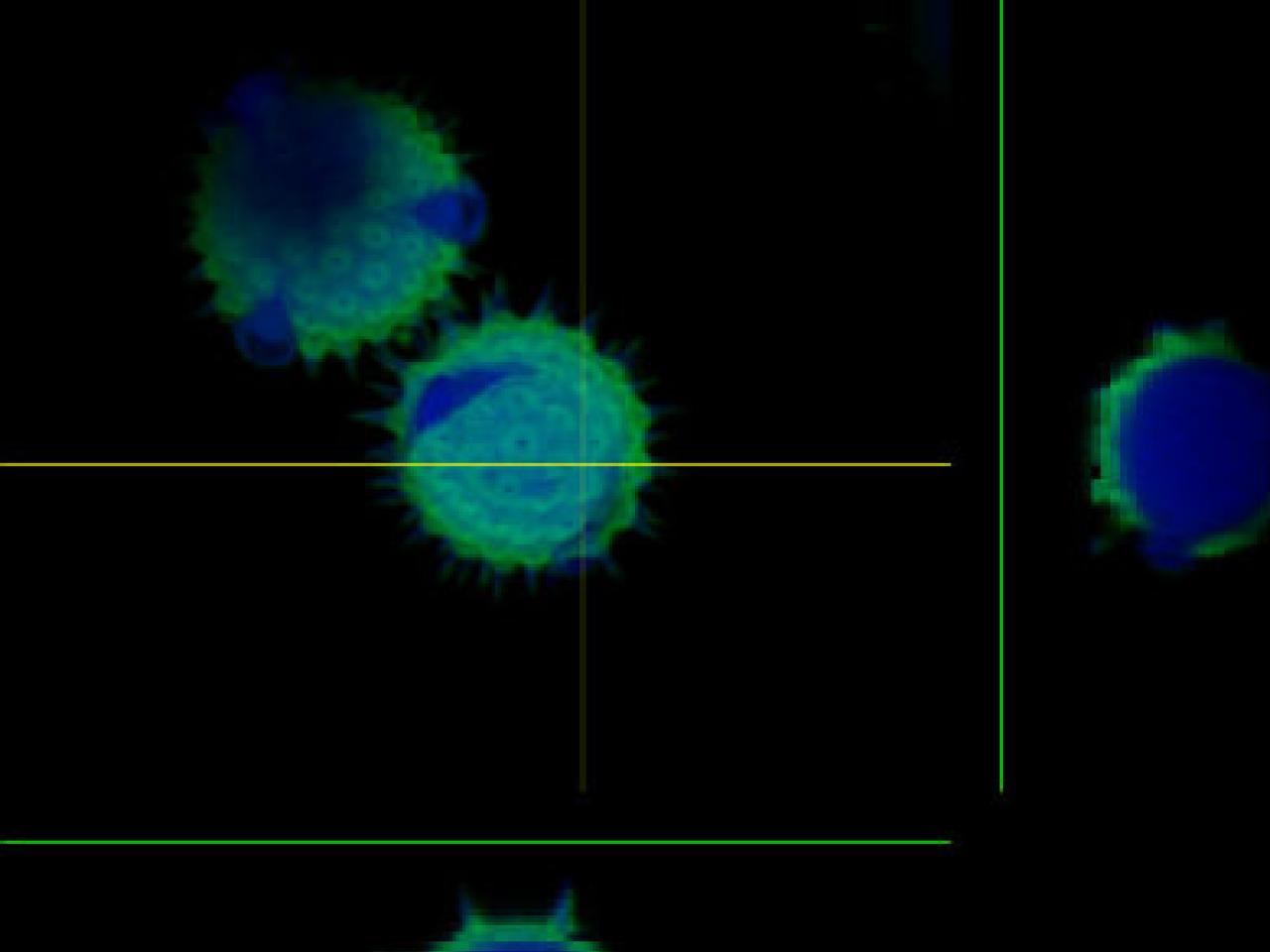
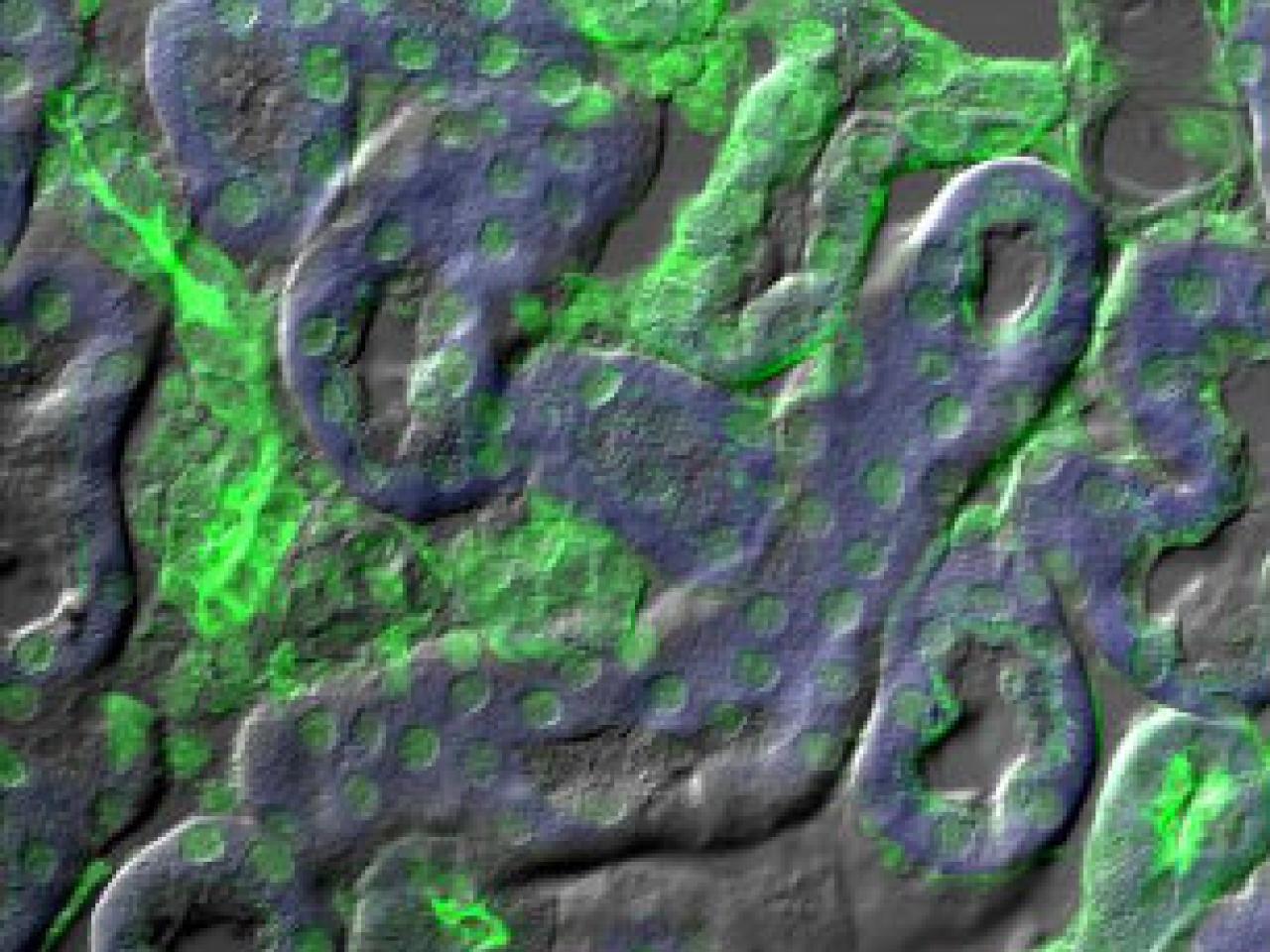
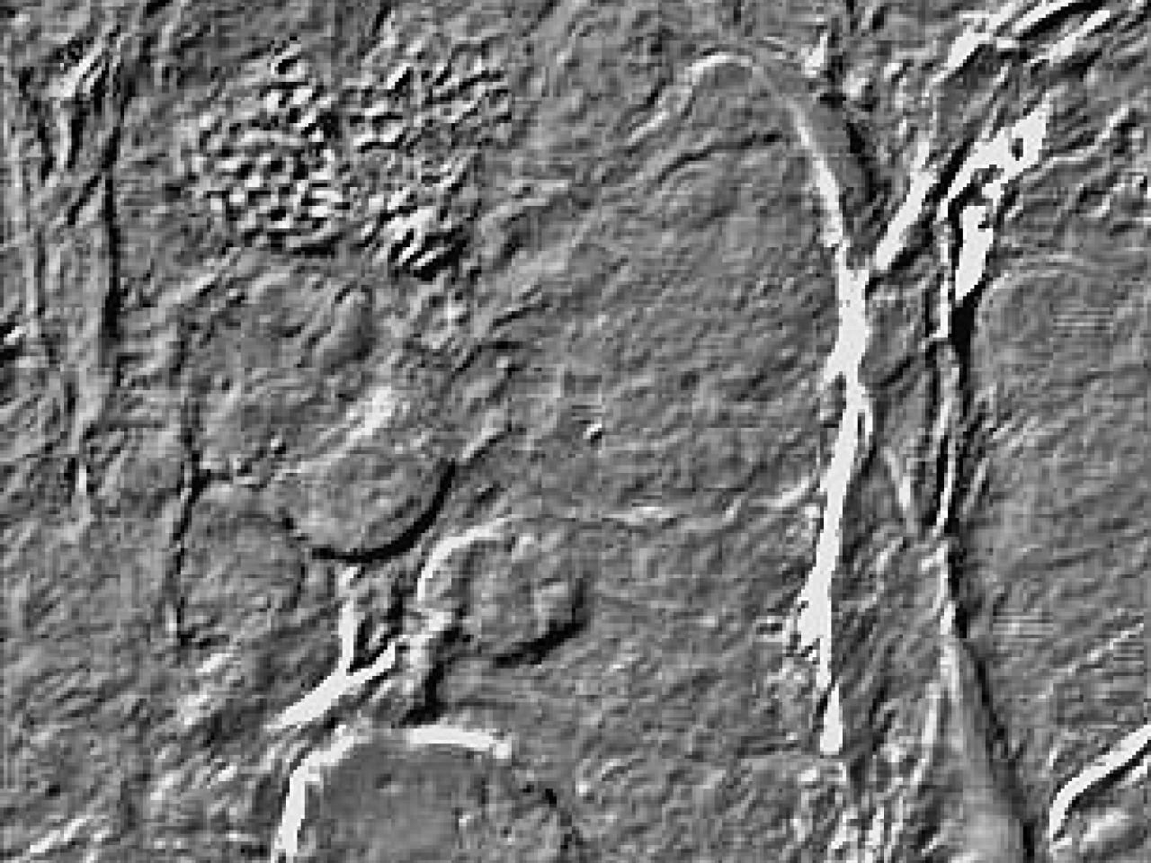
These microscopy images showcase a variety of advanced imaging techniques used in cell and tissue biology research. From multicolor fluorescence images highlighting specific cellular components like cytoskeletons, mitochondria, and nuclei, to sophisticated surface topology maps and differential interference contrast views, each image reveals different aspects of cellular architecture and organization. The collection demonstrates how modern microscopy can visualize both labeled and unlabeled biological structures, providing researchers with detailed views of everything from individual cellular components to complex tissue networks in both two and three dimensions.