Evident Scientific FV1000MPE Multiphoton Laser Scanning Confocal

Multiphoton microscope system enabling deep tissue imaging including label-free collagen detection. Located in BRT 264A.

System Overview

The Evident Scientific (formerly Olympus) FV1000MPE Multiphoton Laser Scanning Confocal has:

  • Multiphoton and confocal modes
  • DeepSee MaiTai titanium-sapphire laser for imaging deep into fixed or living tissues.
  • Second harmonic generation (SHG) imaging - SHG can image collagen, myosin, and some polysaccharides such as cellulose and starch without staining.  
  • A vibratome is available for thick sectioning of fixed tissues.
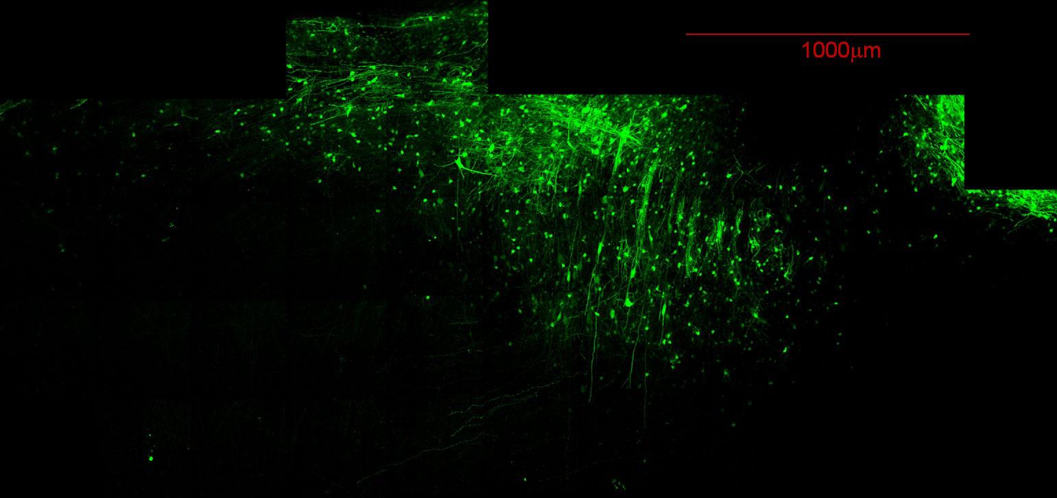
  • A motorized stage is available for capturing tiled images or tiled image stacks.

Access and Support

The Evident Scientific FV1000MPE Multiphoton is available for independent use. Complete confocal microscope (single photon) training is required with NO exception. Training is performed on an individual basis using the researchers samples and instrument rates and technical assistance fees apply during individual instruction.

BSL-2 training from Ohio State EHS is required for all researchers wishing to perform live cell imaging at the CMIF. A signed form must be submitted to the facility before the start of any live cell imaging project.

Tiled image stacks of GFP-expressing neurons in an ‘optically cleared’ rat brain section.
A fluorescence microscopy image showing neurons labeled in bright green against a black background. The image reveals an extensive network of neuronal processes and cell bodies, with long axons extending vertically through the field of view. A red scale bar in the top right indicates 1000μm. The image appears to be a tiled or stitched composite showing a large field of view of neural tissue.