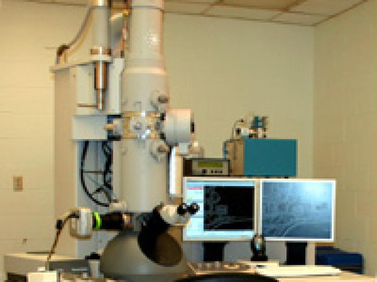
Transmission Electron Microscope for Biological Research at Ohio State's CMIF. Located in BRT 245G. This microscope has been DECOMMISSIONED and is no longer available for use.
Transmission Electron Microscope
FEI Tecnai G2 Spirit BioTwin TEM (80kV).
High contrast polepiece and low kV operating voltage, ideal for imaging biological samples
Room temperature samples only
Full service TEM sample preparation and imaging available upon request
Microscope
FEI Tecnai G2 Biotwin
High Tension
80 kV
Magnification
22-300,000X magnification
Optical System
4 Megapixel XR40 Optronics CCD camera
AMT Image Capture Software
Resolution
0.34 nm (line resolution)
Stage Control
Ambient (20° C) operating temperature
Single tilt sample holder with alpha tilt ± 80°
Note
For other electron microscopy services (e.g., aberration-corrected high-resolution imaging, chemical analysis, cryo-electron microscopy), please contact the Center for Electron Microscopy and Analysis.
Heading
Sample Images
Gallery Image
Carousel Description
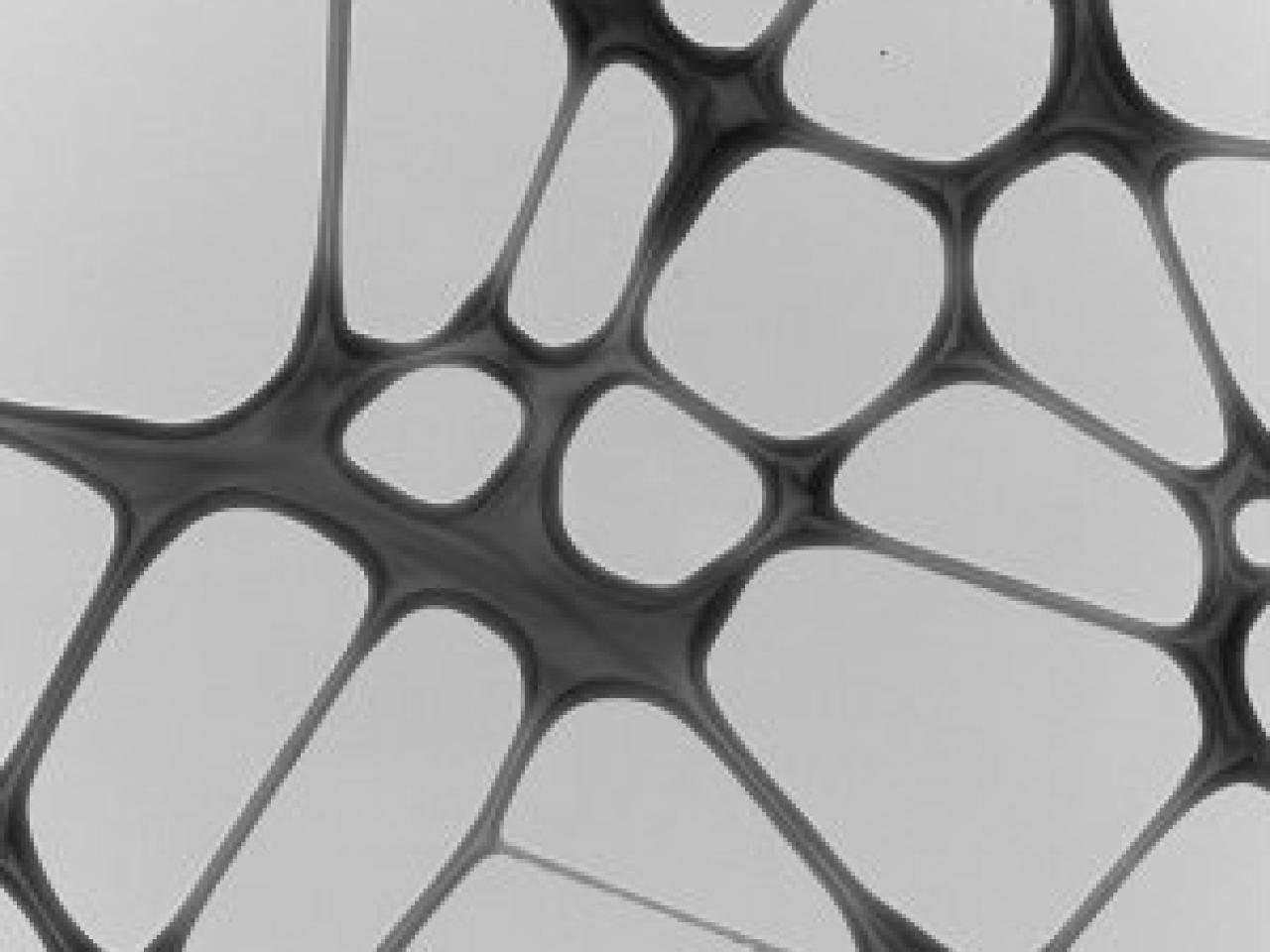
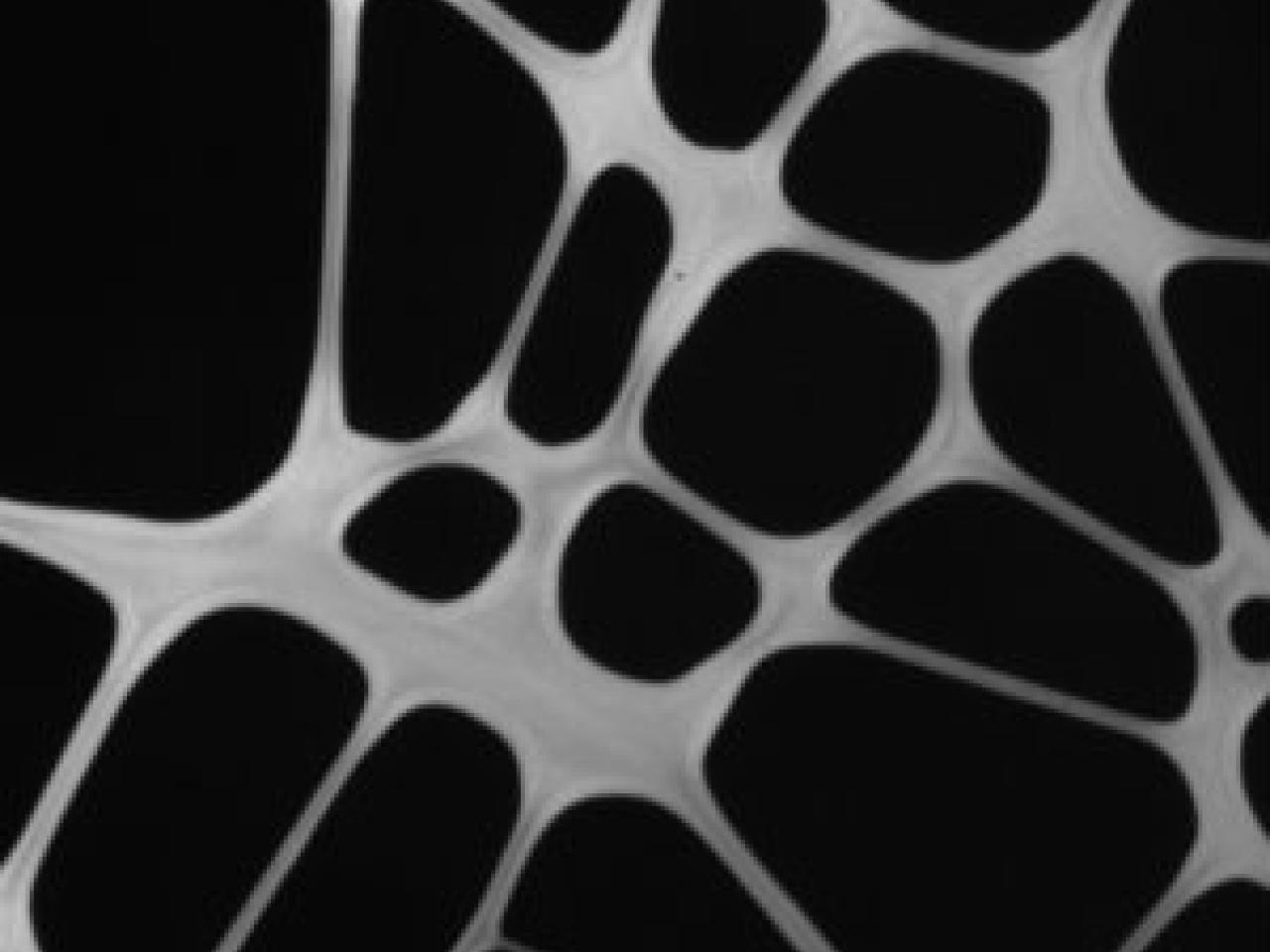
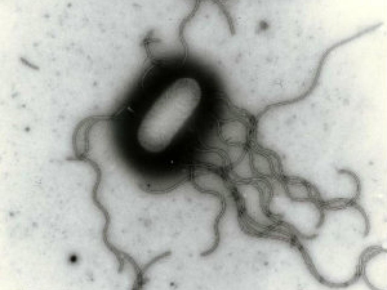
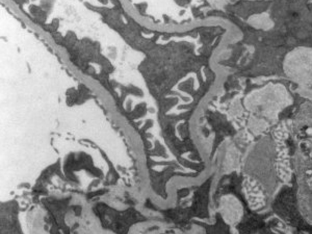
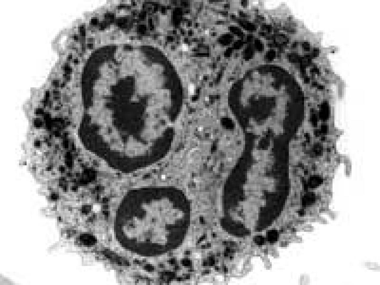
These transmission electron microscope (TEM) images showcase various biological structures at extremely high magnification, demonstrating the power and versatility of electron microscopy in biological research. The collection includes views of different cellular and subcellular structures: a bacterial cell with its characteristic flagella, a polymorphonuclear white blood cell with its distinctive multi-lobed nucleus, complex membrane structures showing cellular ultrastructure, and what appears to be plant or tissue cross-sections showing honeycomb-like patterns in both positive and negative contrast. The first image shows the TEM instrument itself - a sophisticated piece of equipment featuring a tall column and dual monitors for digital image acquisition and analysis. Together, these images illustrate how TEM enables researchers to visualize biological structures at the nanoscale level, revealing details that would be impossible to see with conventional light microscopy.