Liquid crystal filter for RGB (color), near infrared, and monochrome imaging
Pixel size, 6.45 x 6.45 um
Pixels, 1.4 MP, 1392 x 1040 pixels
Stage Control
Manual stage
Heading
Sample Images
Gallery Image
Carousel Description
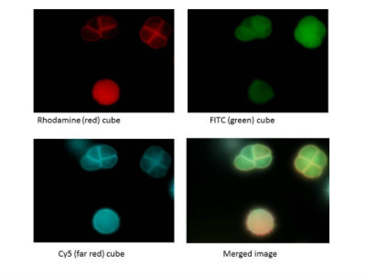
The image suite demonstrates the versatility of fluorescence microscopy techniques in visualizing cellular structures and molecular targets. The first image showcases actin filament organization, combining three fluorescent channels to reveal cytoskeletal architecture (green), nuclear positioning (blue), and an additional cellular marker (red). This is followed by a series of four related panels demonstrating multichannel imaging capabilities using different fluorescent markers - Rhodamine (red), FITC (green), and Cy5 (displayed in cyan) - with the fourth panel showing how these channels can be merged to create a composite view. This type of imaging approach allows researchers to simultaneously visualize multiple cellular components or molecular targets within the same sample, providing valuable insights into their spatial relationships and potential interactions. The clarity and specificity of the fluorescent labeling, combined with the high-contrast dark background, enables detailed examination of cellular structures and their organization within the tissue or culture environment.